


 大包装询价
大包装询价 产品介绍 评论(0)
宿主来源
Rabbit抗原名称
Caldesmon分子别名
CDM, CALD1, CAD免疫原
Synthetic Peptide细胞定位
Intracellular, CytoskeletonAccession
Q05682克隆号
SDT-056-48抗体类型
Rabbit mAb应用
ICFCM, IHC-P, ICC, WB, IP反应种属 ?
Hu, Ms, Rt纯化方式
Protein A浓度
0.5mg/ml标记
Unconjugated性状
Liquid缓冲体系
PBS, 40% Glycerol, 0.05%BSA, 0.03% Proclin 300储存条件
12 months from date of receipt / reconstitution, -20 °C as supplied
| 应用 | 稀释度 |
|---|---|
| ICFCM | 1:500 |
| WB | 1:1000 |
| IHC-P | 1:2000 |
| IP | 1:25 |
| ICC | 1:500 |
Caldesmon is a binding protein of smooth actin and calmodulin, a cytoskeleton-associated protein present in thin filaments (smooth and striated muscles) that regulates the interaction of actin and myosin. It is available in low molecular weight (l-Caldesmon) and high molecular weight (h-Caldesmon). The latter is thought to be present only in visceral and vascular smooth muscle and myoepithelium. Caldesmon is commonly used in the diagnosis of smooth muscle differentiation tumors.
免疫印迹
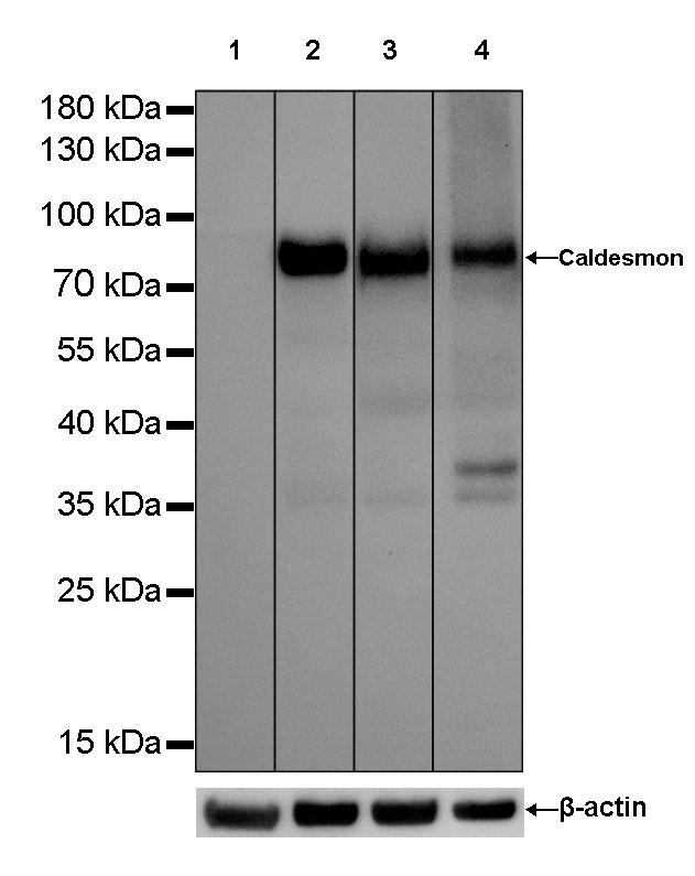
WB result of Caldesmon Rabbit mAb
Primary antibody: Caldesmon Rabbit mAb at 1/1000 dilution
Lane 1: MCF7 whole cell lysate 20 µg
Lane 2: Hela whole cell lysate 20 µg
Lane 3: MDA-MB-231 whole cell lysate 20 µg
Lane 4: HT-1080 whole cell lysate 20 µg
Negative control: MCF7 whole cell lysateSecondary antibody: Goat Anti-Rabbit IgG, (H+L), HRP conjugated at 1/10000 dilution
Predicted MW: 80 kDa
Observed MW: 80 kDa
Exposure time: Lane 1 and lane 2 : 5s
Lane 3 and lane 4 : 10s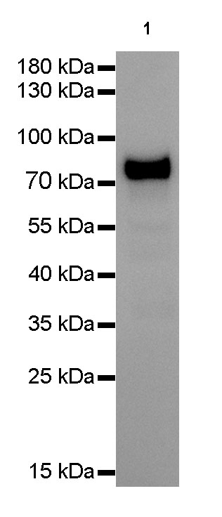
WB result of Caldesmon Rabbit mAb
Primary antibody: Caldesmon Rabbit mAb at 1/1000 dilution
Lane 1: NIH/3T3 whole cell lysate 20 µgSecondary antibody: Goat Anti-Rabbit IgG, (H+L), HRP conjugated at 1/10000 dilution
Predicted MW: 80 kDa
Observed MW: 80 kDa
Exposure time: 5s
流式分析
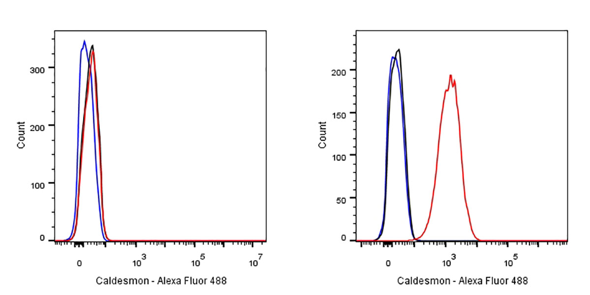
Flow cytometric analysis of MCF7 (left) / HeLa (right) cells labelling Caldesmon antibody at 1/500 (0.1ug) dilution/ (red) compared with a Rabbit monoclonal IgG (Black) isotype control and an unlabelled control (cells without incubation with primary antibody and secondary antibody) (Blue). Goat Anti-Rabbit IgG Alexa Fluor® 488 was used as the secondary antibody. Negative control: MCF7
免疫沉淀
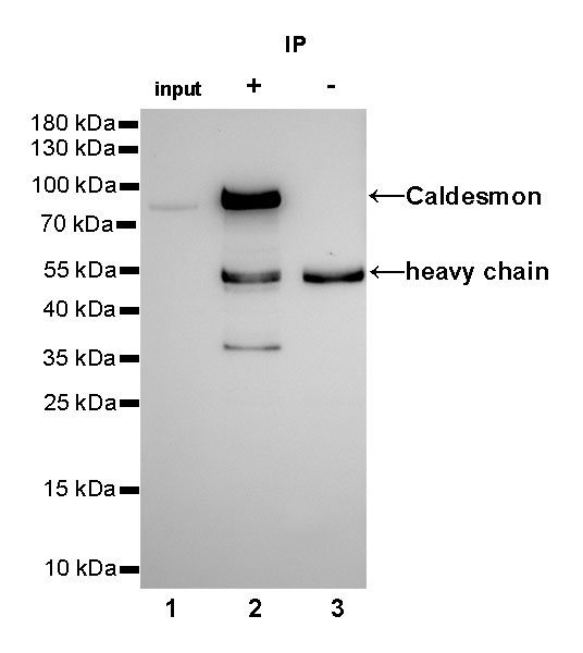
Caldesmon Rabbit mAb at 1/25 dilution (2µg) immunoprecipitating Caldesmon in 0.4mg HeLa whole cell lysate.
Western blot was performed on the immunoprecipitate using Caldesmon Rabbit mAb at 1/1000 dilution.
Secondary antibody (HRP) for IP was used at 1/400 dilution.
Lane 1 : HeLa whole cell lysate 10µg (input)
Lane 2 : Caldesmon Rabbit mAb IP in HeLa whole cell lysate
Lane 3 : Rabbit monoclonal IgG IP in HeLa whole cell lysate
Predicted MW: 80 kDa
Observed MW: 80 kDa
Exposure time: 60s
免疫组化
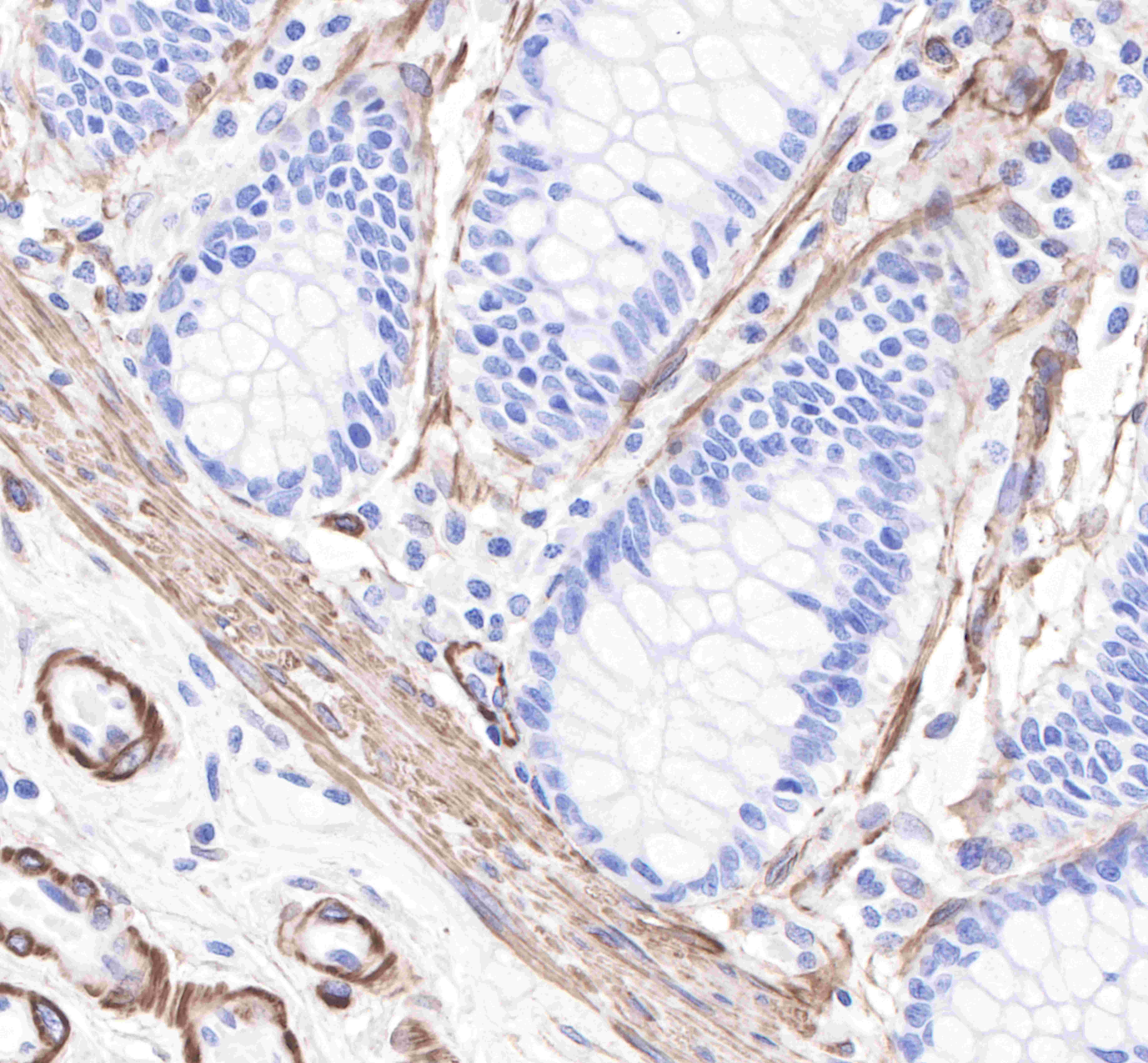
IHC shows positive staining in paraffin-embedded human colon.
Anti-Caldesmon antibody was used at 1/2000 dilution, followed by a Goat Anti-Rabbit IgG H&L (HRP) ready to use. Counterstained with hematoxylin.
Heat mediated antigen retrieval with Tris/EDTA buffer pH9.0 was performed before commencing with IHC staining protocol.
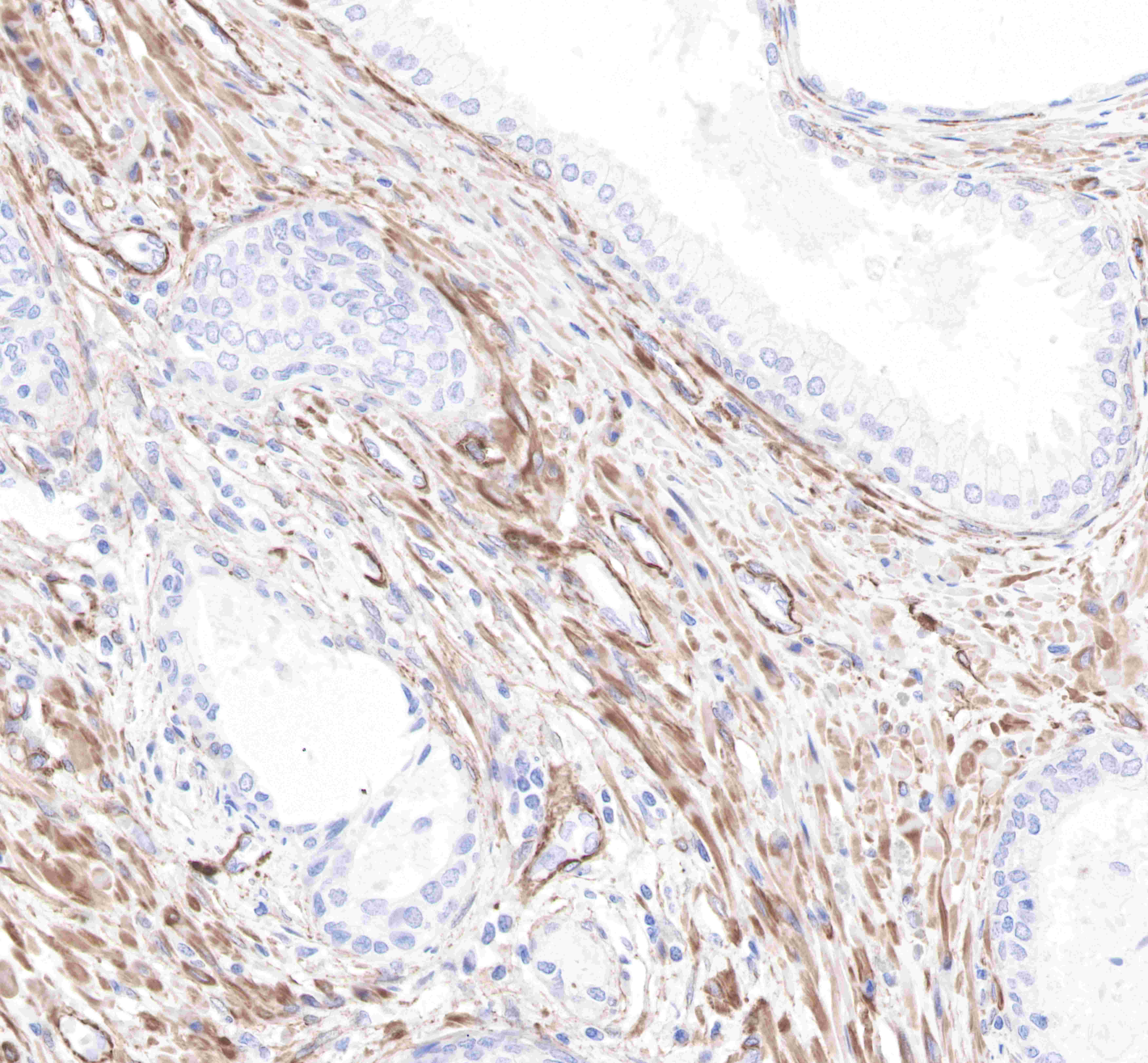
IHC shows positive staining in paraffin-embedded human prostate.
Anti-Caldesmon antibody was used at 1/2000 dilution, followed by a Goat Anti-Rabbit IgG H&L (HRP) ready to use. Counterstained with hematoxylin.
Heat mediated antigen retrieval with Tris/EDTA buffer pH9.0 was performed before commencing with IHC staining protocol.
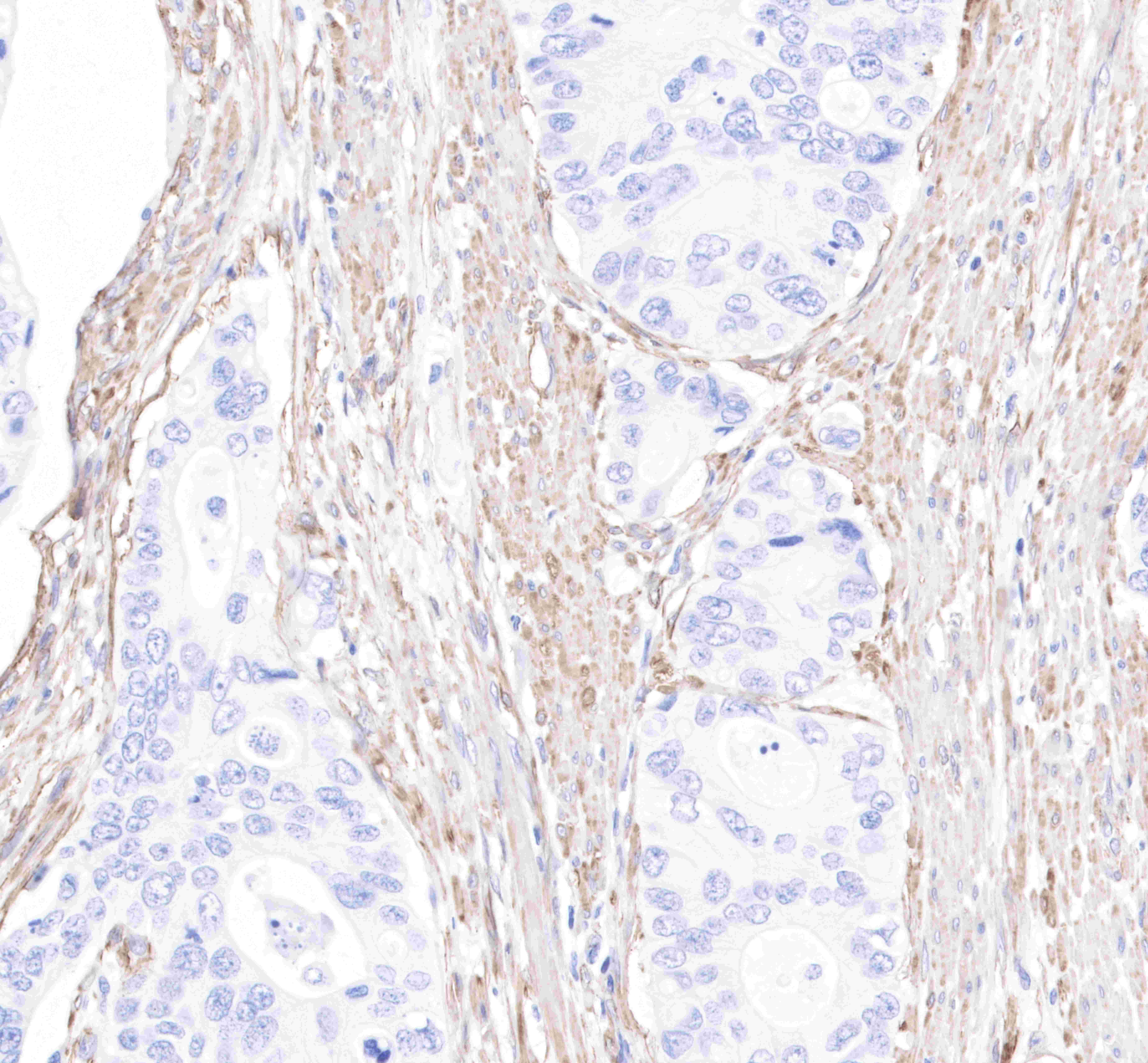
IHC shows positive staining in paraffin-embedded human colon cancer. Anti-Caldesmon antibody was used at 1/2000 dilution, followed by a Goat Anti-Rabbit IgG H&L (HRP) ready to use. Counterstained with hematoxylin.
Heat mediated antigen retrieval with Tris/EDTA buffer pH9.0 was performed before commencing with IHC staining protocol.
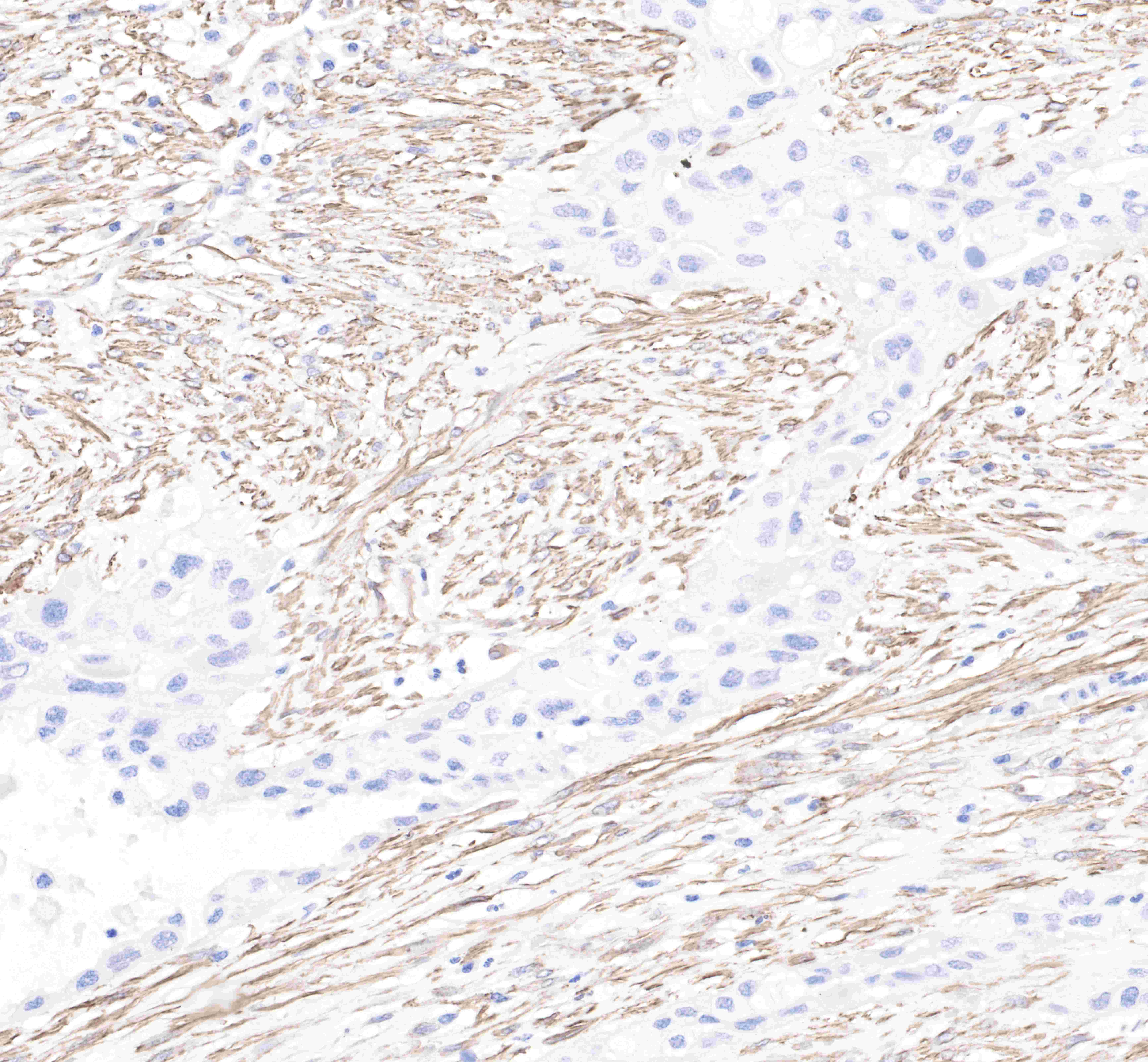
IHC shows positive staining in paraffin-embedded human pancreas cancer.
Anti-Caldesmon antibody was used at 1/2000 dilution, followed by a Goat Anti-Rabbit IgG H&L (HRP) ready to use. Counterstained with hematoxylin.
Heat mediated antigen retrieval with Tris/EDTA buffer pH9.0 was performed before commencing with IHC staining protocol.

IHC shows positive staining in paraffin-embedded mouse kidney.
Anti-Caldesmon antibody was used at 1/2000 dilution, followed by a Goat Anti-Rabbit IgG H&L (HRP) ready to use. Counterstained with hematoxylin.
Heat mediated antigen retrieval with Tris/EDTA buffer pH9.0 was performed before commencing with IHC staining protocol.
IHC shows positive staining in paraffin-embedded rat kidney.
Anti-Caldesmon antibody was used at 1/2000 dilution, followed by a Goat Anti-Rabbit IgG H&L (HRP) ready to use. Counterstained with hematoxylin.
Heat mediated antigen retrieval with Tris/EDTA buffer pH9.0 was performed before commencing with IHC staining protocol.
Negative control.IHC shows negativee staining in paraffin-embedded human skelteal muscle.
Anti-Caldesmon antibody was used at 1/2000 dilution, followed by a Goat Anti-Rabbit IgG H&L (HRP) ready to use. Counterstained with hematoxylin.
Heat mediated antigen retrieval with Tris/EDTA buffer pH9.0 was performed before commencing with IHC staining protocol.
免疫细胞化学
ICC shows positive staining in HeLa cells (top panel) and negative staining in MCF7 cells (below panel). Anti-Caldesmon antibody was used at 1/500 dilution (Green) and incubated overnight at 4°C. Goat polyclonal Antibody to Rabbit IgG - H&L (Alexa Fluor® 488) was used as secondary antibody at 1/1000 dilution. The cells were fixed with 4% PFA and permeabilized with 0.1% PBS-Triton X-100. Nuclei were counterstained with DAPI (Blue). Counterstain with tubulin (Red).


评论(0)